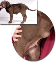
Hernias are very common in human medicine, especially in males. Hernias in general are a weakness or opening within a muscle mass that allows other tissues to pass through. In men, they are usually inguinal hernias which are found in the groin area where there is a tiny natural opening within a band of muscle. When a hernia occurs here, the opening enlarges and the intestines from the abdominal area pass through it, producing a swelling immediately under the skin. Types of Hernias in Dogs:  In dogs, there are also hernias involving the muscles that surround the abdomen and they are commonly found at two locations. The first site would be in the groin area on the inner surface of the rear leg - an inguinal hernia. In dogs, there are also hernias involving the muscles that surround the abdomen and they are commonly found at two locations. The first site would be in the groin area on the inner surface of the rear leg - an inguinal hernia.
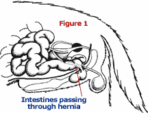
The second site would be the "belly button" where the umbilical cord had connected the puppy to its mother. A hernia at this location is called an umbilical hernia. In both cases, abdominal organs such as the intestines or fat pass through the opening and lie just beneath the skin. Another common hernia site in dogs involves the internal muscle that separates the abdomen and chest. That muscle is called the diaphragm, so the hernia is therefore referred to as a diaphragmatic hernia. The intestines and other abdominal organs (such as liver and stomach) are able to pass through the opening within the diaphragm into the chest cavity. There they take up a portion of the space normally occupied by the lungs. A hernia is, therefore, usually nothing more than an abnormal opening in a muscle through which other tissues of the body pass. Consequences of Hernias: The idea that a section of intestine or other structure might slip through one of these openings and move under the skin or into a different body cavity (such as the chest) doesn't seem like a big problem. However, in many cases, a hernia that goes untreated can have a fatal outcome. Usually, the problems that occur are not caused by the intestines or other organs being in an abnormal position or from the displacement of other tissues that are supposed to be there. Rather, in most instances, a problem arises when the blood supply of the herniated tissues is affected.  Figure #1 shows a hernia that involves the abdominal wall and a section of the small intestine. A portion of the intestine has slipped through a small hole in the muscular wall. This is exactly how an umbilical hernia appears. Figure #1 shows a hernia that involves the abdominal wall and a section of the small intestine. A portion of the intestine has slipped through a small hole in the muscular wall. This is exactly how an umbilical hernia appears. Notice the stricture (abnormal narrowing) of the intestine itself. This could easily prevent the passage of food through this section of the intestine, effectively causing an obstruction or blockage. This would certainly lead to the death of the animal if it were not treated. 
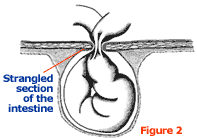
More importantly however, please look at Figure #2. This shows a close up of the intestinal wall as it passes through the hernia. Notice how the blood vessels are twisted and constricted. Blood will not be able to flow back and forth from between this portion of the intestine and the rest of the body. It means that the section of intestine that has passed through the hole in the abdominal wall will lose its blood supply. It will be deprived of oxygen and nutrients and when this occurs, it dies. Symptoms: The symptoms associated with a hernia like the one pictured in Figure 1 and 2 may initially relate to the inability of food to pass through this constricted section of intestine. Muscles within the wall of the intestine are responsible for moving food and water through the organ. Waves of contractions called peristalsis propel the contents along the length of the intestine. When an obstruction is encountered like the one described, the peristaltic waves reverse direction and move the food backward through the entire digestive tract. This results in food and water being vomited. After this portion of the tract has emptied, the animal usually goes off food and refuses to eat. They may still drink water because liquids might be able to pass through the restricted section of the intestine or be absorbed prior to that point. Once the blood vessels are affected, however, the clinical signs change drastically. The area will become swollen and painful. Without adequate oxygen and nutrients, the intestinal tissues initially develop cramps just like your leg does when you cross it and it "goes to sleep". And if the flow of blood is completely lost, cell death occurs. The pain then becomes severe. The animal will probably develop a fever, become lethargic, and go completely off food and water. As these tissues break down, the toxins from bacteria that normally live in the intestine make their way into the rest of the animal’s body. As the tissue dies, the affected area turns into an abscess and many different harmful metabolic waste products are flushed throughout the animal’s body. All of these substances (bacterial toxins and metabolic waste products) seriously affect the various organ systems of the body. Liver and/or kidney failure are quite common in these situations. Without treatment, the animal will usually die within 24 to 48 hours. As an owner, do not take a hernia in your pet lightly. In many cases, they are disasters just waiting to happen. Do not buy a puppy that has a hernia unless you have a veterinarian examine it so you will understand what treatment is necessary and the potential cost. Some hernias found in young dogs can wait for repair until the time they are spayed or neutered. In older dogs, we generally repair a hernia as soon as possible once they are discovered. Treatment: Hernias are repaired by replacing the herniated (displaced) structures back into their correct position and then suturing closed the abnormal openings. This often requires the use of specialized techniques and long-lasting suture material. Hereditary Potential: As a note, umbilical hernias in puppies are a genetic or congenital defect in over 90% of the cases. The disorder is passed from generation to generation just like the color of the coat or the animal’s overall size. Very, very rarely are they caused by trauma or excessive pressures during whelping. Animals that have a hernia or had a surgical repair of a hernia should never be used for breeding. Additionally, those adults that produce puppies with this condition should not be bred again. 

|  Free Forum Hosting