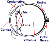
 The eye is a very delicate, yet surprisingly durable organ. It consists of several layers. The cornea is a transparent layer that covers the front of the eye. The iris is the colored part of the eye and it is responsible for letting in more or less light. The lens gathers and 'bends' light in order to focus it on the retina.
In between the cornea and lens is an area of fluid which bathes the lens and helps it focus. The retina lines the inside of the eye and converts light into signals which travel down the optic nerve to the brain.
A large area between the lens and the retina contains a jelly-like fluid called 'vitreous.' The vitreous gives the eye its form and shape, provides nutrients, and removes waste products.
The retina
The retina is the structure affected in PRA. This important part of the eye receives the light gathered and focused by the other eye structures. It takes the light and essentially converts it into electrical nerve signals that the brain, via the optic nerve, interprets as vision. The retina contains photoreceptors, called rods and cones, which help the animal see in darkness (rods) and see certain colors (cones).
What is PRA?
There are multiple forms of PRA which differ in the age of onset and rate of progression of the disease. Some breeds experience an earlier onset than others; other breeds do not develop PRA until later in life.
Normally, the photoreceptors in the retinas develop after birth to about 8 weeks of age. The retinas of dogs with PRA either have arrested development (retinal dysplasia) or early degeneration of the photoreceptors.
Retinal dysplastic dogs are usually affected within two months of birth and may be completely blind by one year. Dogs with retinal degeneration are affected from one year to eight years of age and the symptoms progress slowly.
PRA worsens over time. The affected animal experiences night blindness initially because the rods are affected first. The condition progresses to failed daytime vision.
What are the signs of PRA?
Signs may vary depending on the type of PRA and its rate of progression. PRA is non painful and outward appearance of the eye is often normal, i.e.; no redness, excess tearing, or squinting.
Owners may notice a change in personality of their dog such as a reluctance to go down stairs or down a dark hallway. This is characteristic of night blindness, in which vision may appear to improve during the daytime.
As the disease progresses, owners can observe a dilation of the pupils and the reflection of light from the back of the eye. If the blindness is progressing slowly, the owner may not notice any signs until the dog is in unfamiliar surroundings and the lack of vision is more apparent. In some animals, the lens of the eyes may become opaque or cloudy.
How is PRA diagnosed?
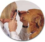 Depending on the form of PRA, characteristic changes in the retina and other parts of the eye may be observed through an ophthalmic examination by a veterinary opthalmologist.
More sophisticated tests such as electroretinography may also be used. Both tests are painless and the animal does not have to be anesthetized. If no abnormalities are found during the exam by a board certified veterinary ophthalmologist, the dog can be certified free of heritable eye disease through the Canine Eye Registration Foundation (CERF).
How is PRA treated?
Unfortunately, there is no treatment for PRA, nor a way to slow the progression of the disease. Animals with PRA usually become blind. Dogs are remarkably adaptable to progressive blindness, and can often seem to perform normally in their customary environments. Evidence of the blindness is more pronounced if the furniture is rearranged or the animals are in unfamiliar surroundings.
Can PRA be prevented?
PRA has been shown to have a genetic component. Puppies from parents who have no history of the disease and have been certified free of PRA will have less risk of developing the disease. Affected animals should not be bred and should be spayed or neutered. The littermates or parents of animals with PRA should also not be bred. If your dog develops PRA, notify the breeder, if possible.
In the last several years, DNA is being used to identify which genes are responsible for PRA. In one form of PRA called 'rod cone dysplasia 1' (rcd1), which affects Irish Setters, the gene mutation has been identified.
Progressive rod-cone degeneration (prcd) is the most widespread form of PRA and affects many breeds including Poodles, American and English Cocker Spaniels, Labrador Retrievers, and Portuguese Water Dogs.
Prcd starts with night blindness and progresses to total blindness at 3 to 5 years of age. The late onset of clinical signs in prcd is particularly devastating to breeding programs because many dogs have already been bred prior to the onset of symptoms. Thus, the development of a genetic test for this disease which could identify both affected animals and those that just carry the gene would be particularly useful.
The researchers at the James A. Baker Institute have found a set of genetic markers that usually indicate the presence of the gene mutation that causes prcd in English Cockers, Labrador Retrievers, Chesapeake Bay Retrievers, and Portuguese Water Dogs. Although the exact gene mutation that causes prcd has not been found, every dog that is affected with prcd has two copies of the genetic marker and every dog that does not have the marker is clear of prcd.
Unfortunately, this test for the genetic marker is not as accurate in diagnosing prcd as it would be if the actual gene mutation was found. This is because dogs that are not affected by prcd may still have the genetic marker present and have false positive test results. Thus dogs that are positive for the gene marker may be either false positives or may be carriers of the disease or may actually have the disease. The marker test may be done at a very early age, so potential breeding animals may be selected when they are still puppies.

 Important Announcement